Radiation Risk for Oral Surgery Imaging
Understanding the radiation risk for oral surgery imaging is an important step in the process of putting a patient’s mind at ease regarding the necessity for such imaging studies. Since 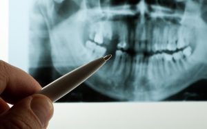 radiographic studies play an integral role in the diagnosis and treatment of many dental and oral conditions, putting patients at ease is imperative. The issue of radiation risk is best understood by identifying why such tests are necessary, the safety of such tests, and how clinicians can minimize any risk to their patients.
Why are Radiographic Exams Necessary?
Most often radiographic dental imaging consists of intraoral or extraoral x-rays or CT scans. Radiographic imaging is an invaluable tool for dentists and oral surgeons for both the diagnosis and treatment of oral diseases. These images provide pictures of the hard and soft tissues of the mouth, and can alert your dentist or oral surgeon to conditions like tooth decay, bone disease, periodontal disease, gum infection, or even tumors.
Well-trained practitioners use criteria guidelines to assist them in making appropriate recommendations for radiographs. It is understandable that patients might worry that doctors are using a one-size-fits-all approach to ordering x rays. It is therefore up to the doctor to justify every radiograph in order to maximize diagnostic data while minimizing risk of radiation exposure.
Are Dental Radiographs Safe?
It is generally reported that dental radiographs are very safe and that the cancer risk from such tests is negligible. When undergoing a dental imagining study, a patient is exposed to far less radiation than what they are exposed to on a typical day in their life from their ordinary environment.
All dentists and oral surgeons are required to maintain an ethical obligation to keep radiation exposure as low as reasonably achievable. This is achieved by using the fastest imagining method possible, using the smallest beam of x-ray to achieve the necessary image, and using the proper exposure and processing techniques to obtain quality images the first time.
How can any Risks be Minimized?
Patients can play an active role in reducing the risk of radiation exposure during dental x rays. The dentist or oral surgeon should have a thorough discussion with the patient regarding their medical and dental history before ordering any x rays.
During the tests, the patient’s body will be shielded from unnecessary radiation with a leaded apron or collar. Staff members performing the tests should have the necessary credentials and skills to produce quality images without needing to do retakes, thereby limiting unnecessary exposure.
For the best in oral surgery care, contact the trusted team at Oral and Maxillofacial Surgery of Utah. Put your mind at ease by allowing us to answer all of your questions regarding any radiation risk for oral surgery imaging.
Comments are closed.